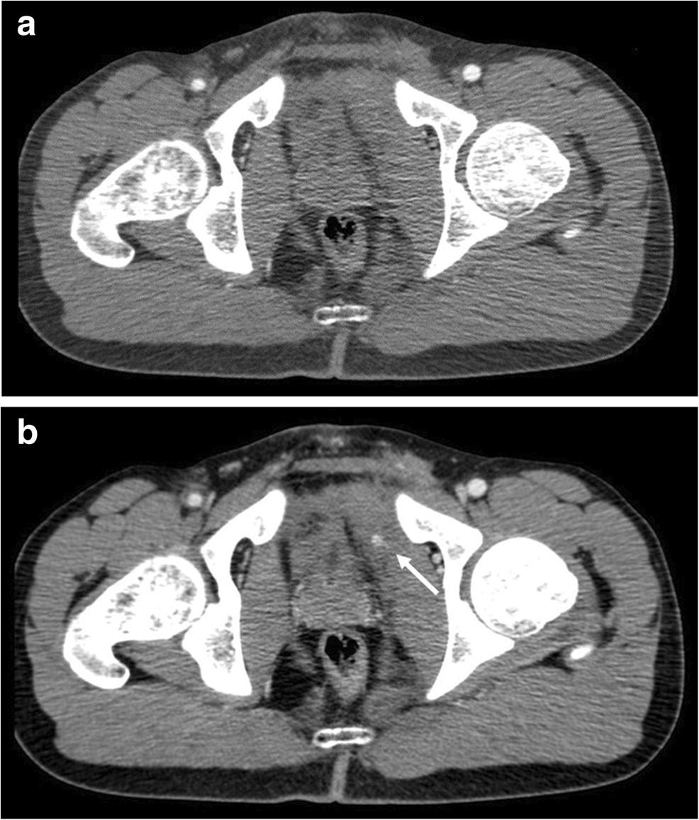
A CT Pelvis Scan (Computed Tomography of the pelvis) is a non-invasive imaging technique that provides detailed cross-sectional images of the pelvic region, including bones, muscles, organs, and blood vessels. It is commonly used to diagnose fractures, tumors, infections, or abnormalities in pelvic structures.
This scan is particularly helpful in assessing conditions such as trauma, cancer staging, vascular disorders, or unexplained pelvic pain. Contrast dye may be used to enhance visualization of blood vessels and soft tissues. The procedure is quick, typically taking 10-30 minutes, and requires the patient to lie still on a motorized table. While the scan involves exposure to ionizing radiation, it is considered safe when medically necessary.
Indications:
1. Pelvic
pain: Evaluate cause of acute or chronic pelvic pain.
2. Trauma:
Assess pelvic injuries, fractures, or bleeding.
3. Cancer
diagnosis: Detect and stage pelvic tumors, such as cervical, ovarian, or
prostate cancer.
4. Infections:
Identify abscesses, pelvic inflammatory disease, or other infections.
5. Urinary
tract problems: Diagnose kidney stones, bladder issues, or ureteral
obstruction.
6. Gynecological
disorders: Evaluate conditions like endometriosis, fibroids, or ovarian
cysts.
7. Prostate
disorders: Diagnose prostate cancer, prostatitis, or benign prostatic
hyperplasia.
8.
Rectal disorders: Evaluate rectal cancer, fistulas, or abscesses.
9. Pre-operative
evaluation: Assess patients before pelvic surgery.
10. Post-operative
evaluation: Monitor healing and detect potential complications after pelvic
surgery.
11. Hematuria:
Investigate blood in the urine.
12. Pelvic
organ prolapse: Evaluate descent of pelvic organs, such as the bladder or
uterus.
13.
Fistula evaluation: Diagnose abnormal connections between pelvic organs or
the skin.
14. Inflammatory
bowel disease: Monitor Crohn's disease or ulcerative colitis in the pelvic
area.
Benefits of
CT Pelvis scan:
1. Accurate
diagnosis: Helps diagnose various pelvic conditions, including cancer,
infections, and injuries.
2. Detailed
images: Provides high-resolution images of pelvic organs, tissues, and
blood vessels.
3. Guides
treatment: Helps guide biopsies, tumor resections, and other surgical
procedures.
4. Monitors
progress: Tracks the effectiveness of treatments and detects potential
complications.
5. Detects
additional findings: May detect other conditions, such as kidney stones or
bowel obstruction.
6. Reduces
uncertainty: Clarifies ambiguous findings from other imaging tests.
7. Supports
research: Contributes to the study of pelvic diseases and conditions.
8. Enhances
patient care: Leads to better diagnosis, treatment, and patient outcomes.
9.
Quick and non-invasive: Typically a fast and painless procedure.
10. Improved
patient safety: Reduces the risk of complications in surgical procedures.
11.
Better understanding: Provides a clear understanding of the extent of
disease.
12. Improved
treatment planning: Helps plan radiation therapy and other treatments.
13.
Detects pelvic fractures: Identifies fractures or dislocations in the
pelvic bones.
14. Evaluates
pelvic organ prolapse: Assesses descent of pelvic organs, such as the
bladder or uterus.
