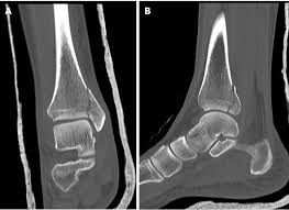
A CT (Computed Tomography) scan of the ankle is a detailed imaging test that uses X-rays and advanced computer processing to create cross-sectional images of the ankle's bones, joints, and soft tissues. It is particularly useful for diagnosing and assessing complex injuries or conditions that may not be fully visible on standard X-rays.
Purpose of a CT Ankle Scan
- Trauma and Fractures: To evaluate complex fractures, dislocations, or small bone fragments in the ankle joint.
- Arthritis: To assess the extent of joint damage in conditions such as osteoarthritis or rheumatoid arthritis.
- Bone Diseases: To detect bone infections, tumors, or abnormalities.
- Pre-Surgical Planning: To provide detailed anatomical information for surgical intervention.
- Congenital Anomalies: To investigate structural issues present from birth.
Procedure
- Preparation: The patient may be asked to remove any metal objects and might be given specific instructions if contrast material is used.
- Positioning: The patient lies on a CT table, and the ankle is positioned within the scanner's field.
- Scanning: The CT machine rotates around the ankle, taking multiple X-ray images, which are processed to form 3D or cross-sectional views.
Advantages
- Provides more detailed images of bones compared to standard X-rays.
- Can detect subtle fractures and assess the alignment of the joint.
- Non-invasive and quick, often taking less than 10 minutes.
Limitations
- Exposure to a small amount of ionizing radiation.
- May require contrast material, which carries a risk of allergic reaction in some patients.
A CT ankle scan is a valuable tool for orthopedic and diagnostic purposes, offering precise visualization of the ankle's internal structures for effective treatment planning.
