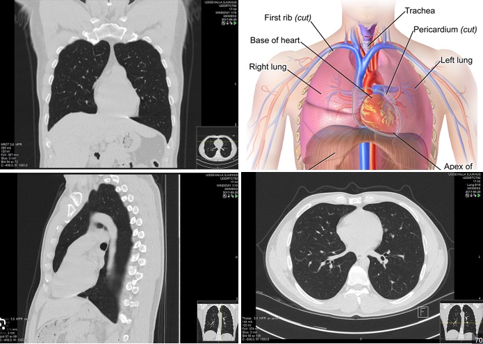
Uses of doing CT Chest Scan

A
chest CT scan is a painless, noninvasive imaging test that uses X-rays to
create detailed pictures of the chest. The procedure involves the following
steps:
CT CHEST INDICATION:
Lung problems:
Infections, lung cancer, blocked blood flow,
pulmonary embolism, and other lung issues
Chest injuries:
Injuries
to the heart, blood vessels, lungs, ribs, and spine
Tumors:
Tumors
that arise in the chest, or tumors that have spread there from other parts of
the body
Blood clots:
Symptoms
that suggest blood clots in the lungs, such as chest pain, rapid breathing, or
shortness
1. Preparation
You
may be asked to change into a hospital gown, remove glasses and metal objects,
and arrive 20 minutes early. You may also need to provide a blood sample to
check kidney function if contrast dye is used.
2. Positioning
You
lie on a narrow table that slides into the center of the scanner. A pillow and
straps hold you in place to prevent movement.
3. Imaging
The
X-ray beam rotates around you, taking many images of your chest from different
angles. You may be asked to hold your breath for a short time.
4. PROCESSING
The
images are sent to a computer, which processes them to create cross-sectional
and 3D images.
5. Results
The
images are displayed on a monitor.
CT
scans can provide more detailed images than regular X-rays, and can help
identify injuries or diseases in the chest. They are useful for emergencies
because they can provide real-time images of internal bleeding.
The
main risk of a CT scan is radiation exposure, but the benefits of the
information obtained usually outweigh the risks. Low-dose CT scanning
techniques can reduce radiation exposure.
Benefits
of a CT [Computed Tomography] chest scan:
Diagnostic Benefits:
1.
Accurate detection of lung nodules, tumors, and cancers.
2.
Evaluation of chest pain, cough, and difficulty breathing.
3.
Diagnosis of pulmonary embolism (blood clots in lungs).
4.
Detection of pneumonia, bronchitis, and other infections.
5.
Assessment of lung damage from injury or trauma.
Patient Benefits:
1. Quick
procedure (typically 5-10 minutes).
2.
Minimal discomfort.
3.
Non-invasive.
4.
No radiation exposure (compared to traditional angiography).
5.
High-resolution images.
Clinical Benefits:
1.
High sensitivity and specificity for detecting lung diseases.
2.
Detailed images of lungs, mediastinum, and surrounding tissues.
3.
Helps identify vascular diseases (e.g., aortic aneurysm).
4.
Useful for monitoring disease progression.
5.
Can reduce need for additional imaging tests.
Some
common indications for a CT chest scan include:
-
Lung cancer screening
-
Chest pain or difficulty breathing
-
Coughing up blood
-
Fever with respiratory symptoms
-
Trauma or injury
-
Pre-operative evaluation
CLINICAL PULMONARY MANIFESTATION OF
COVID-19 infections and the radiography procedures used for diagnosis:
Clinical Pulmonary Manifestations:
1.
Pneumonia
2.
Acute Respiratory Distress Syndrome (ARDS)
3.
Bronchiolitis
4.
Pulmonary Embolism
5.
Pleural Effusion
*Radiography Procedures:
Computed
Tomography (CT) Scan:
1.
More sensitive than CXR for early detection
2.
High-resolution CT (HRCT) for detailed evaluation
3.
Findings:
- GGO
- Consolidation
- Crazy-paving pattern
- Reverse halo sign
- Pulmonary embolism
CT Scan Protocols:
1.
Non-contrast CT
2.
Thin-section CT (1-2 mm)
3.
High-resolution CT (HRCT)
4.
Reconstruction algorithms (e.g., lung window)
Radiological Features:
Early Stage (0-4 days):
1.
Unilateral or bilateral GGO
2.
Peripheral distribution
3.
Lower lobe predominance
Progressive Stage (5-10 days):
1.
Consolidation
2.
Increased opacity
3.
Bilateral involvement
Severe Stage (11+ days):
1.
Extensive consolidation
2.
Pulmonary cavitation
3.
Pneumothorax
Other Imaging Modalities:
1.
MRI (limited role)
2.
PET-CT (research purposes)
Imaging Recommendations:
1.
American College of Radiology (ACR) guidelines
2.
Fleischner Society guidelines
3.
WHO guidelines
Important Notes:
1.
Imaging findings may lag behind clinical symptoms
2.
Radiography should be used in conjunction with clinical evaluation and
laboratory tests
3.
Repeat imaging may be necessary to monitor disease progression
For a doing painless CT CHEST Scan in Chennai, consider Madras Scans & Labs as your go-to destination. It is the most trusted scan center in Chennai with its exceptional and secure diagnostic services at an affordable pricing system.
Give us a quick call at 9514400800 or visit us at https://www.madrasscan.in to know more about other scans and our attractive price package.
